A whole new world under the microscope
Business insights

Business insights
While many of his contemporaries used what were then newly developed optical instruments such as telescopes to observe distant heavenly bodies, one man explored the unknown right under his nose. Dutch draper Antoni van Leeuwenhoek is regarded as the father of microbiology. His home-made microscopes allowed him to reach levels of magnification surpassing anything that had gone before.

In the Netherlands, the 17th century is referred to as the ‘Golden Age’ to this day. International trade was flourishing, cities and ports were growing, and the economic boom was accompanied by a cultural zenith. Freedom of religion attracted numerous writers and scholars to the country, where they could teach and publish their works unhindered. In this period of prosperity and liberal ideas, Antoni van Leeuwenhoek (1632–1723) was born in Delft to a basket-maker and a brewer’s daughter. He developed an early interest in mathematics and physics. At his mother’s request, however, he went to Amsterdam at the age of 16 to learn the trade of a draper. Six years later he returned to his hometown, where he bought a house, married and opened a draper’s shop.

The oldest surviving drawing of a van Leeuwenhoek microscope from 1753. All the microscopes van Leeuwenhoek left to the Royal Society (a fellowship of scholars founded in Britain in 1660 to promote excellence in science) were of this type. The element facing the eye can be seen at the top; the fixture for mounting specimens is at the bottom.
Drapery was big business in the Netherlands at the time, so the young van Leeuwenhoek’s economic prospects could not have been better. Moreover, a clever and reliable manner earned him considerable respect and opened the door to important official positions over the years. He served as chamberlain for the Delft assembly chamber, for example, and was later appointed as a land surveyor and even became the city’s official ‘wine gauger’. Van Leeuwenhoek took advantage of the resultant early financial independence for the passionate pursuit of his favourite hobby: microscopy. “It probably all began when he wanted to build a thread counter – a kind of magnifying glass to examine the density and quality of woven fabrics,” explains Dr. Timo Mappes, Professor of the History of Physics at Jena’s Friedrich Schiller University and Founding Director of the Deutsches Optisches Museum (German Museum of Optics) in the same city. “What he was able to see with this small instrument must have fascinated him so much that, from this time on, he developed a genuine passion for the subject.”
Telescopes and microscopes in the 17th century were mostly made with two or more lenses. Since production methods could not yet make glass that was sufficiently homogeneous, the images were usually distorted and often also had coloured edges that were clearly discernible. Van Leeuwenhoek therefore developed a device that comprised only one spherical lens enclosed between two metal plates. He devoted a great deal of attention to polishing lenses, perfecting a technique that ultimately allowed him to magnify objects up to 270 times at least. These lenses far exceeded the capabilities of previous microscopes and gave him an insight into a world no one before him had ever seen. “It was he who first sketched bacteria and described many other microorganisms,” Mappes points out. “So he did the groundwork that attracted the attention of subsequent generations to this topic and finally allowed them to understand the issues involved.”
Antoni van Leeuwenhoek was a talented draughtsman who used this skill to keep a record of his findings for posterity. This figure shows a drawing of spermatozoans from various mammals from the year 1677. This spectacular discovery by van Leeuwenhoek proved the similarity between the spermatozoans of different mammals.
Dated 1719, this picture shows the blood corpuscles that van Leeuwenhoek saw contained in blood vessels.

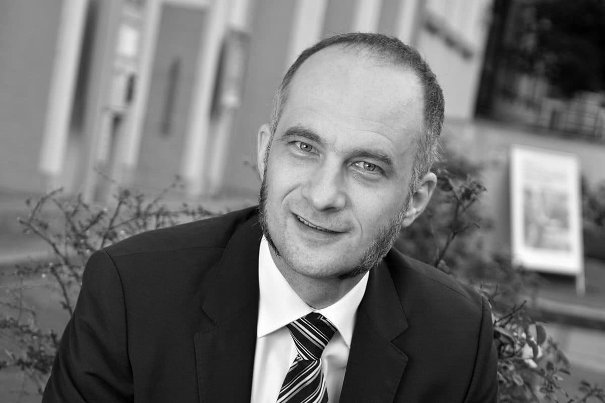
Professor Timo Mappes (45) graduated in mechanical engineering at the University of Karlsruhe in 2003. He then earned his doctorate here with an experimental thesis on microstructure technology. In 2006 he was a member of the core team that brought the University of Karlsruhe, the Karlsruhe Research Centre and the Karlsruhe Institute of Technology (KIT) together. Following on from visiting professorships in Denmark and France, Mappes moved to Carl Zeiss AG in 2012. Here, he led global research and development in eyeglass lenses, staffed by a good 200 employees on four continents. He was also responsible for the company’s IP portfolio. In mid-2018 he was appointed Professor of the History of Physics at the Friedrich Schiller University in Jena, where he also serves as Founding Director of the German Museum of Optics (D.O.M.). Over the past two decades, Mappes’ personal interest in the documentation and scientific history of microscope construction has earned him an outstanding reputation via the medium of various forms of publication.
A layman, van Leeuwenhoek enjoyed no formal scientific education. Dutch was the only language he spoke. Yet his work earned him considerable international respect. He sent several hundred letters detailing his observations to the Royal Society, a London-based fellowship of scholars, for example. The Society translated his notes into Latin and even elected him as a member in 1680. At the same time, he also corresponded with many prominent individuals of his day, some of whom – including Czar Peter the Great, Queen Mary II of England and Gottfried Wilhelm Leibniz – visited him in Delft.
Throughout his life, the self-educated scientist kept the way he produced his equipment secret. Why he did this is still not known. What is certain, however, is that it took a lot more practice to use his microscopes than it does to work with modern models. “The metal frame was held up to the eye, which peered through a hole less than two millimetres across. The spherical lens was held in place behind this hole,” Professor Mappes explains. “The object to be examined was placed on the point of a needle on the other side, with a thread allowing the needle to be moved like a screw. A second screw varied the distance from the lens to bring the object into focus.” Since van Leeuwenhoek evidently built a separate device for every kind of specimen, there is evidence that his workshop brought forth more than 500 microscopes for different applications.
After initially investigating anything and everything he could put in front of the lens, the draper-turned-scientist gradually began to focus more selectively on natural science, keeping a conscientious written record of what he saw. The doctrine of spontaneous generation postulated that certain small living creatures could emerge from dust and was still a widely held theory at the time. Van Leeuwenhoek suspected that this was wrong, however, and shared his insights. He was the first person to describe the uncontrolled movements of sperm and red blood corpuscles. Another of his spectacular observations was the finding that the bacteria he found in his dental plaque and called ‘little creatures’ stopped moving when he treated them with vinegar. “There are many interrelationships that he did not yet understand,” Mappes admits. “But his empirical approach enabled him to recognise that there is no end to what can be discovered in the realms of the small. And that is what he went off in search of.”


Less than a dozen of van Leeuwenhoek’s microscopes have survived to this day. Four of them are at the Museum Boerhaave in Leiden, two are at the Deutsches Museum in Munich. The remainder are in other museums or are privately owned. “It took a hundred years before devices with a comparable resolution could be made and his observations could be reproduced on a large scale,” Professor Mappes stresses. One could accuse the Dutchman of not keeping more detailed and extensive records of how he produced his microscopes, the professor concedes. “But the way in which he discovered and described the microcosmos in his immediate vicinity was outstanding.”
Connecting Specialists to Pioneering Projects